Category: Blog

Your blog category

  • Regaining Function After Injury: The Role of Tendon Transfer Surgery

    Regaining Function After Injury: The Role of Tendon Transfer Surgery

    Hand Tendon Transfer Surgery in Malleshwaram

    Tendon transfer surgery is a specialized procedure aimed at restoring hand function lost due to nerve injuries, muscle damage, or congenital conditions. By redirecting functioning tendons to compensate for impaired ones, this surgery can significantly improve mobility and strength in the hand. At Sapiens Clinic in Malleshwaram, we offer expert evaluation and surgical solutions to help patients regain optimal hand function.

    What Is Tendon Transfer Surgery?

    Tendon transfer involves relocating a healthy tendon from its original attachment to a new one to restore lost function. This procedure is particularly beneficial when specific muscles or tendons are non-functional due to:

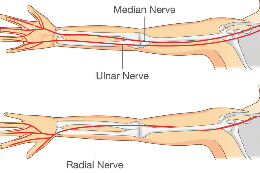
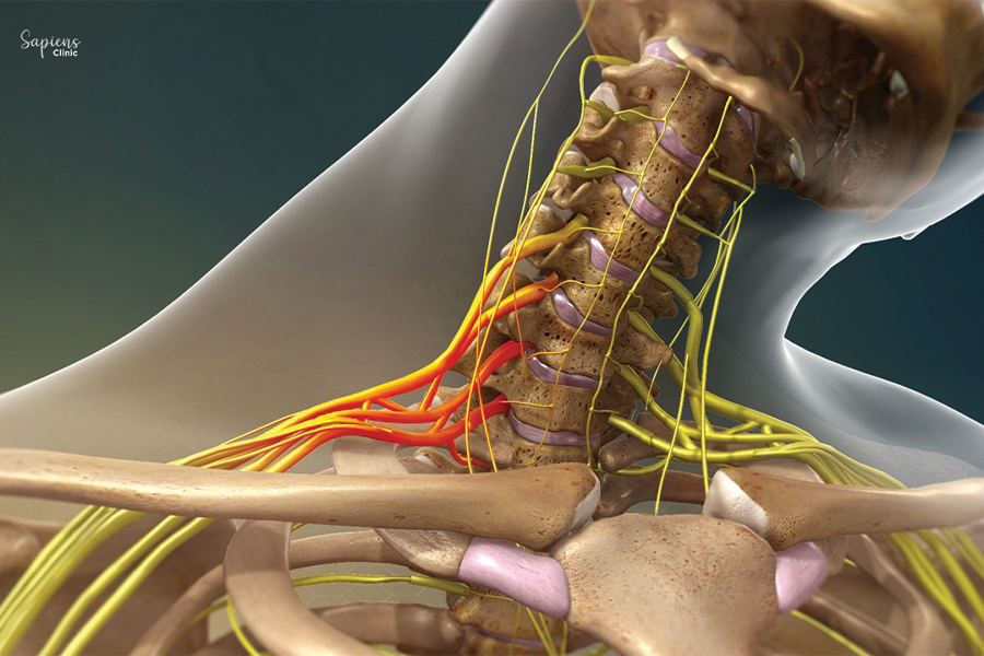
    • Peripheral nerve injuries (e.g., radial, median, or ulnar nerve palsies)
    • Spinal cord injuries leading to muscle paralysis
    • Brachial plexus injuries
    • Congenital hand deformities
    • Tendon ruptures or lacerations

    The goal is to restore specific movements such as wrist extension, finger flexion, or thumb opposition by rerouting tendons from muscles that have retained their function.

    Indications for Hand Tendon Transfer

    Tendon transfer surgery may be considered in cases where:

    • Nerve repair is not feasible or has failed
    • Muscle function cannot be restored through other means
    • There is a need to correct muscle imbalances or deformities
    • Rehabilitation alone has not achieved desired functional outcomes

    Common conditions treated include:

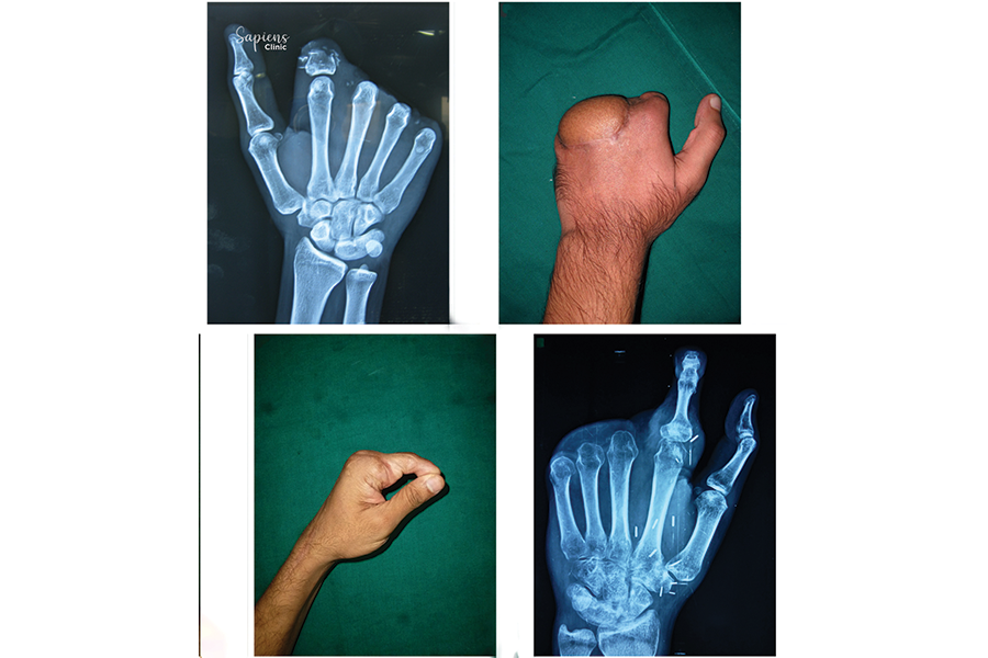
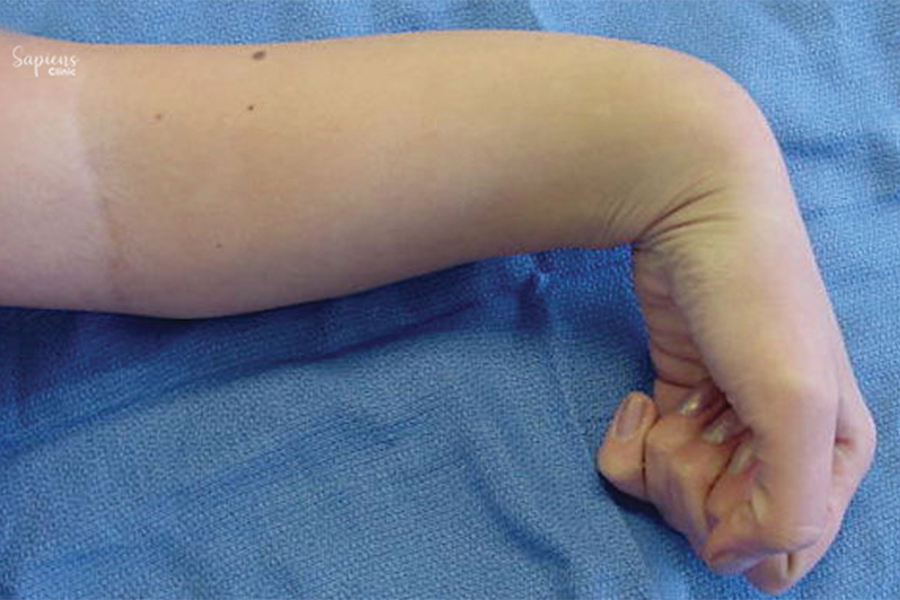
    • Radial nerve palsy (wrist drop)
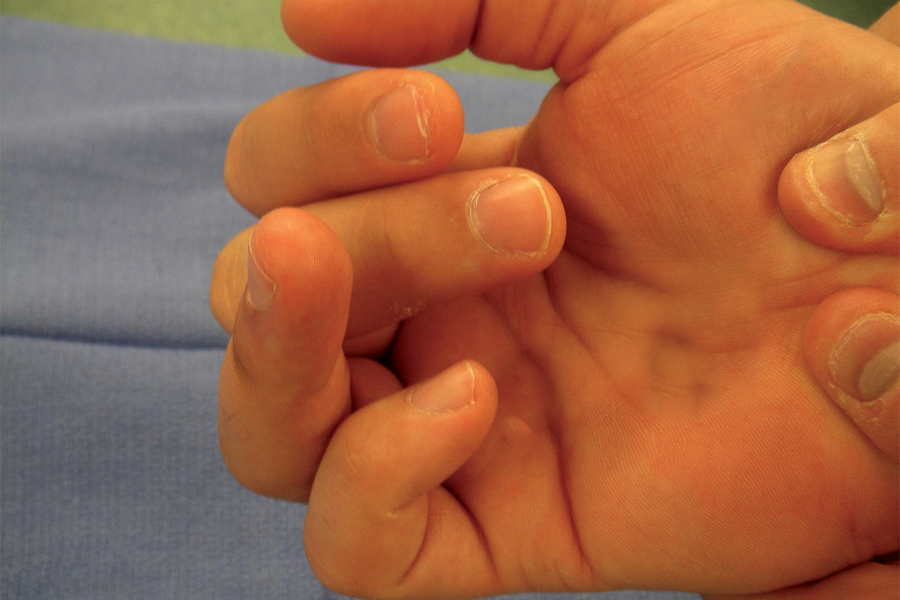
    • Ulnar nerve palsy (claw hand)
    • Median nerve palsy (loss of thumb opposition)
    • Cerebral palsy-related hand dysfunction
    • Post-traumatic tendon or nerve injuries

    Surgical Procedure

    The procedure involves:

    1. Assessment: Identifying functional muscles and tendons suitable for transfer.
    2. Planning: Determining the specific tendons to be rerouted to restore desired movements.
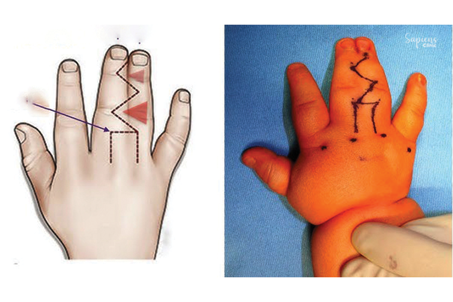
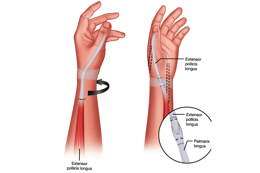
    3. Surgery: Detaching the selected tendon from its original insertion and reattaching it to a new site to perform the lost function.
    4. Stabilization: Securing the transferred tendon and immobilizing the hand to facilitate healing.

    The choice of donor tendon depends on factors such as muscle strength, range of motion, and the specific functional deficit being addressed.

    Recovery and Rehabilitation

    Postoperative care is crucial for successful outcomes:

    • Immobilization: The hand is typically immobilized in a splint or cast for several weeks to allow healing.
    • Physical Therapy: Rehabilitation begins with gentle movements, progressing to strengthening exercises to retrain the transferred tendon for its new function.
    • Duration: Full recovery and return to daily activities may take several months, depending on the complexity of the surgery and individual healing rates.

    Benefits of Tendon Transfer Surgery

    The procedure aims to:

    • Restore lost hand and finger movements
    • Improve grip strength and dexterity
    • Enhance the ability to perform daily tasks
    • Correct deformities and improve hand appearance
    • Reduce pain and discomfort associated with muscle imbalances

    Risks and Considerations

    As with any surgical procedure, tendon transfer carries potential risks:

    • Infection at the surgical site
    • Scar formation affecting tendon gliding
    • Incomplete restoration of function
    • Need for additional surgeries in some cases

    A thorough preoperative evaluation and adherence to postoperative rehabilitation protocols are essential to minimize these risks.

    Conclusion

    Tendon transfer surgery offers a viable solution for restoring hand function when other treatments have failed or are not possible. If you’re experiencing hand dysfunction due to nerve or tendon injuries, consult Dr. Darshan Kumar A. Jain, Orthopedic Specialist at Sapiens Clinic, Malleshwaram, for a comprehensive evaluation and personalized treatment plan.

    FAQs

    1. How long does recovery take after tendon transfer surgery?

    Recovery time varies but typically spans several months, including immobilization and rehabilitation phases.

    2. Will I regain full function of my hand?

    While the goal is to restore as much function as possible, outcomes depend on various factors, including the extent of the injury and adherence to rehabilitation.

    3. Are there alternatives to tendon transfer surgery?

    Alternatives may include nerve repair, physical therapy, or the use of orthotic devices, depending on the specific condition.

    4. Is the procedure painful?

    Pain is managed with appropriate anesthesia during surgery and medications postoperatively.

    5. What is the success rate of tendon transfer surgeries?

    Success rates are generally high, especially when performed by experienced surgeons and followed by dedicated rehabilitation.

  • Understanding Joint Transfer Procedures: When and How They Are Performed

    Understanding Joint Transfer Procedures: When and How They Are Performed

    Joint Transfer Surgery in Malleshwaram

    Joint transfer surgery is a specialized orthopedic procedure aimed at restoring mobility and function in joints severely damaged by trauma, arthritis, or congenital anomalies. At Sapiens Clinic in Malleshwaram, we offer advanced joint transfer techniques to help patients regain movement and improve their quality of life.

    Understanding Joint Transfer Surgery

    Joint transfer involves relocating a joint from one part of the body to another to replace a non-functional or absent joint. This procedure is particularly beneficial when prosthetic joint replacement is not feasible or has failed. By transferring a patient’s own joint, we aim to achieve better integration and functionality.

    Indications for Joint Transfer

    Joint transfer surgery may be considered in the following scenarios:

    • Traumatic Joint Loss: Severe injuries leading to the loss of joint structures.
    • Failed Joint Replacements: Previous prosthetic implants that have failed or caused complications.
    • Congenital Joint Absence: Birth defects resulting in missing or underdeveloped joints.
    • Infection-Induced Joint Damage: Severe infections leading to joint destruction.

    Each case is evaluated individually to determine the suitability of joint transfer as a treatment option.

    Surgical Procedure

    The joint transfer procedure involves several critical steps:

    1. Donor Joint Selection: Identifying a suitable joint for transfer, often from a less critical area.
    2. Harvesting the Joint: Careful extraction of the donor joint along with its associated structures.
    3. Recipient Site Preparation: Preparing the damaged area to receive the new joint.
    4. Microsurgical Attachment: Connecting bones, tendons, ligaments, and blood vessels to ensure functionality.
    5. Postoperative Care: Monitoring for complications and initiating rehabilitation.

    Advanced imaging and surgical planning are utilized to enhance precision and outcomes.

    Recovery and Rehabilitation

    Recovery from joint transfer surgery involves a comprehensive rehabilitation program :

    • Immobilization: Initial period to allow for healing and integration.
    • Physical Therapy: Customized exercises to restore range of motion and strength.
    • Regular Monitoring: Follow-up visits to assess progress and address any issues.
    • Long-Term Care: Ongoing support to maintain joint function and prevent complications.

    The recovery timeline varies depending on the joint involved and the individual patient’s condition.

    Conclusion

    Joint transfer surgery offers a viable solution for patients with complex joint issues where traditional treatments have failed. At Sapiens Clinic, Malleshwaram, our multidisciplinary teamis dedicated to providing personalized care, utilizing cutting-edge techniques to restore mobility and enhance the quality of life for our patients.

    FAQs

    1. What is the success rate of joint transfer surgeries?

    Success rates vary based on the joint involved and the patient’s overall health, but many patients experience significant improvements in function and pain relief.

    2. Are there risks associated with joint transfer surgery?

    As with any surgical procedure, risks include infection, blood clots, and issues with joint integration. Our team takes all necessary precautions to minimize these risks.

    3. How long is the hospital stay after joint transfer surgery?

    Hospital stays typically range from a few days to a week, depending on the complexity of the surgery and the patient’s recovery progress.

    4. Will I need physical therapy after the surgery?

    Yes, physical therapy is a crucial component of recovery, helping to restore movement and strength in the affected joint.

    5. Can joint transfer surgery be performed on children?

    Yes, with careful consideration, joint transfer surgery can be performed on pediatric patients, especially in cases of congenital joint absence or severe trauma.

  • How Toe Transfer Surgery Can Help Restore Hand Function After Injury

    How Toe Transfer Surgery Can Help Restore Hand Function After Injury

    Toe Transfer Surgery in Malleshwaram

    Toe transfer surgery, also known as toe-to-hand transfer, is a specialized microsurgical procedure aimed at restoring the function and appearance of hands affected by trauma, congenital anomalies, or disease. At Sapiens Clinic in Malleshwaram, we offer advanced toe transfer procedures to help patients regain essential hand functions and improve their quality of life.

    Understanding Toe Transfer Surgery

    Toe transfer surgery involves transplanting a toe (commonly the second toe) to the hand to reconstruct a missing or non-functional finger or thumb. This procedure is particularly beneficial for patients who have lost a digit due to injury or have congenital conditions affecting hand development. The transferred toe provides both aesthetic and functional restoration, enabling patients to perform tasks that require fine motor skills.

    Indications for Toe Transfer

    Toe transfer surgery is indicated in various scenarios, including :

    • Traumatic Amputations: Loss of fingers or thumbs due to accidents or injuries.
    • Congenital Anomalies: Conditions like thumb hypoplasia where the thumb is underdeveloped or absent.
    • Failed Replantation: When previous attempts to reattach a severed digit have been unsuccessful.
    • Tumor Resections: Removal of digits due to cancerous growths.

    In cases where pollicization (reconstruction of the thumb using the index finger) is not feasible or desired, toe transfer offers an effective alternative.

    Surgical Procedure

    The toe transfer procedure is performed under general anesthesia and involves several key steps:

    1. Donor Site Preparation: The selected toe, usually the second toe, is carefully dissected along with its blood vessels, nerves and tendons.
    2. Recipient Site Preparation: The hand is prepared to receive the toe, ensuring proper alignment and integration.
    3. Microsurgical Transfer: Using a microscope, surgeons connect the blood vessels and nerves of the toe to those in the hand to restore circulation and sensation.
    4. Tendon and Bone Fixation: Tendons are attached to facilitate movement, and bones are fixed using pins or plates for stability.

    The procedure requires meticulous planning and execution to ensure optimal functional and aesthetic outcomes.

    Recovery and Rehabilitation

    Postoperative care is crucial for the success of toe transfer surgery :

    • Hospital Stay: Patients typically stay in the hospital for several days to monitor the viability of the transferred toe.
    • Immobilization: The hand is immobilized to allow for proper healing of the surgical connections.
    • Physical Therapy: Rehabilitation begins a few weeks after surgery to restore movement, strength, and dexterity.
    • Follow-Up Visits: Regular check-ups are scheduled to assess healing progress and address any complications.

    Full recovery and functional integration of the transferred toe can take several months, depending on individual healing rates and adherence to rehabilitation protocols.

    Conclusion

    Toe transfer surgery offers a viable solution for individuals seeking to restore hand function and appearance after digit loss or congenital anomalies. At Sapiens Clinic, Malleshwaram, our experienced surgical team employs state-of-the-art microsurgical techniques to deliver personalized care and optimal outcomes for our patients.

    FAQs

    1. Will the loss of a toe affect my ability to walk?

    Most patients adapt well after the removal of a toe, especially the second toe, with minimal impact on walking or balance.

    2. How long does the surgery take?

    The procedure typically lasts between 6 to 12 hours, depending on the complexity of the case.

    3. What are the risks associated with toe transfer surgery?

    As with any surgical procedure, risks include infection, blood vessel or nerve complications, and partial or complete failure of the transferred toe.

    4. Can children undergo toe transfer surgery?

    Yes, children with congenital hand anomalies can benefit from toe transfer surgery, with considerations made for growth and development.

    5. Will the transferred toe look like a finger?

    While the transferred toe may differ slightly in appearance, surgical techniques aim to achieve a natural look and functional integration with the hand.

  • Understanding Spastic Hand Deformity: Managing Muscle Tightness and Function

    Understanding Spastic Hand Deformity: Managing Muscle Tightness and Function

    Spasticity and Upper Limb Deformities Treatment in Malleshwaram

    daily activities, affecting quality of life. At Sapiens Clinic in Malleshwaram, we specialize in diagnosing and treating these conditions, aiming to restore function, reduce discomfort, and enhance overall well-being.

    Understanding Spasticity and Upper Limb Deformities

    Spasticity refers to involuntary muscle stiffness or tightness, often resulting from neurological conditions such as stroke, cerebral palsy, or spinal cord injuries. This increased muscle tone can lead to deformities in the upper limbs, including abnormal postures, joint contractures, and impaired movement patterns.

    Causes and Risk Factors

    Spasticity and resultant deformities can arise from various factors :

    • Neurological Conditions: Such as stroke, traumatic brain injury, cerebral palsy, and multiple sclerosis.
    • Spinal Cord Injuries: Leading to disrupted nerve signals and muscle control.
    • Brain Injuries: Affecting motor control centers.
    • Neurodegenerative Diseases: Progressive conditions impacting nerve function.

    Early identification and management are crucial to prevent long-term complications.

    Symptoms

    Common signs indicating spasticity and upper limb deformities include :

    • Muscle Stiffness: Persistent tightness in the arms or hands.
    • Involuntary Muscle Spasms: Sudden, uncontrolled movements.
    • Abnormal Postures: Such as flexed elbows or wrists.
    • Joint Contractures: Permanent tightening of muscles or tendons.
    • Difficulty with Fine Motor Skills: Challenges in tasks like writing or buttoning clothes.

    Prompt medical evaluation is essential for optimal recovery.

    Diagnosis

    Accurate diagnosis involves:

    • Clinical Evaluation: Assessing muscle tone, reflexes, and movement patterns.
    • Imaging Studies: MRI or CT scans to identify underlying neurological issues.
    • Electrodiagnostic Tests: EMG and nerve conduction studies to evaluate nerve and muscle function.

    These assessments help determine the extent and location of the impairment.

    Treatment Options

    Management strategies are tailored based on the specific condition:

    • Conservative Therapies :
      • Physical Therapy: Exercises to improve flexibility and strength.
      • Occupational Therapy: Training to perform daily activities safely.
      • Medications: Muscle relaxants or antispastic agents.
      • Splinting and Bracing: To maintain joint position and prevent contractures.
    • Interventional Treatments:
      • Botulinum Toxin Injections: To reduce muscle overactivity.
      • Intrathecal Baclofen Therapy: Delivery of medication directly to the spinal fluid.
    • Surgical Interventions:
      • Tendon Release or Transfer: To correct deformities and improve function.
      • Joint Stabilization Procedures: To maintain proper alignment.
      • Selective Dorsal Rhizotomy: Cutting nerve fibers to reduce spasticity.

    Early intervention often leads to better outcomes.

    Recovery and Rehabilitation

    Post-treatment rehabilitation focuses on :

    • Restoring Function: Through targeted exercises and therapies.
    • Pain Management: Utilizing medications and therapeutic modalities.
    • Monitoring Progress: Regular follow-ups to assess recovery and adjust treatment plans.

    Recovery timelines vary based on the severity of the condition and the treatment approach.

    Conclusion

    Spasticity and upper limb deformities can significantly impact daily activities and overall well-being. At Sapiens Clinic, Malleshwaram, our multidisciplinary team is dedicated to providing comprehensive care, from accurate diagnosis to personalized treatment plans, ensuring optimal recovery and prevention of future complications.

    FAQs

    1. What causes spasticity in the upper limbs?

    Spasticity often results from neurological conditions like stroke, cerebral palsy, or spinal cord injuries that disrupt normal muscle control.

    2. Can spasticity be cured?

    While spasticity may not be entirely curable, various treatments can effectively manage symptoms and improve function.

    3. How does botulinum toxin help in treating spasticity?

    Botulinum toxin injections relax overactive muscles, reducing stiffness and improving movement.

    4. Is surgery always necessary for upper limb deformities?

    Not always. Surgery is considered when conservative treatments are ineffective or when deformities significantly impair function.

    5. How long does rehabilitation take after treatment?

    Rehabilitation duration varies depending on the individual’s condition and response to therapy, ranging from weeks to months.

  • Injured a Nerve in Your Arm? What You Need to Know About Upper Limb Nerve Damage

    Injured a Nerve in Your Arm? What You Need to Know About Upper Limb Nerve Damage

    Radial, Ulnar and Median Nerve Palsy Treatment in Malleshwaram

    Nerve palsies affecting the radial, ulnar, and median nerves can significantly impair hand and arm function, leading to challenges in daily activities. At Sapiens Clinic in Malleshwaram, we specialize in diagnosing and treating these conditions, aiming to restore mobility, reduce pain, and enhance quality of life.

    Understanding Nerve Palsies

    The radial, ulnar, and median nerves are crucial for motor and sensory functions in the upper limb. Damage or compression to these nerves can result in palsies, characterized by muscle weakness, loss of sensation, or both.

    Causes and Risk Factors

    Nerve palsies can arise from various factors :

    • Radial Nerve Palsy: Often due to humeral fractures, prolonged compression (e.g., “Saturday night palsy”), or improper use of crutches.
    • Ulnar Nerve Palsy: Commonly results from cubital tunnel syndrome, where the ulnar nerve is compressed at the elbow.
    • Median Nerve Palsy: Typically associated with carpal tunnel syndrome, caused by pressure on the median nerve at the wrist.

    Risk factors include repetitive motions, trauma, anatomical variations, and certain medical conditions like diabetes.

    Symptoms

    Symptoms vary depending on the affected nerve:

    • Radial Nerve Palsy: Wrist drop, inability to extend the wrist and fingers, and numbness on the back of the hand.
    • Ulnar Nerve Palsy: Numbness and tingling in the ring and little fingers, hand weakness, and muscle wasting in severe cases.
    • Median Nerve Palsy: Numbness and tingling in the thumb, index, middle, and part of the ring finger; weakness in thumb opposition.

    Early recognition of these symptoms is crucial for effective management.

    Diagnosis

    Accurate diagnosis involves :

    • Clinical Evaluation: Assessing medical history and physical examination.
    • Electrodiagnostic Tests: EMG and nerve conduction studies to evaluate nerve function.
    • Imaging Studies: MRI or ultrasound to visualize nerve structures and identify compression points.

    These assessments help determine the extent and location of nerve involvement.

    Treatment Options

    Management strategies are tailored based on the specific condition :

    • Conservative Therapies:
      • Physical Therapy: Exercises to maintain joint mobility and muscle strength.
      • Occupational Therapy: Training to perform daily activities safely.
      • Medications: Pain relievers or anti-inflammatory drugs.
      • Splinting: Immobilization to prevent further nerve irritation.
    • Surgical Interventions:
      • Nerve Decompression: Relieving pressure on compressed nerves.
      • Nerve Repair or Grafting: Reconnecting or replacing damaged nerve segments.
      • Tendon Transfers: Redirecting tendons to restore function.

    Early intervention often leads to better outcomes.

    Recovery and Rehabilitation

    Post-treatment rehabilitation focuses on :

    • Restoring Function: Through targeted exercises and therapies.
    • Pain Management: Utilizing medications and therapeutic modalities.
    • Monitoring Progress: Regular follow-ups to assess recovery and adjust treatment plans.

    Recovery timelines vary based on the severity of the injury and the treatment approach.

    Conclusion

    Radial, ulnar and median nerve palsies can significantly impact daily activities and overall well-being. At Sapiens Clinic, Malleshwaram, our multidisciplinary team is dedicated to providing comprehensive care, from accurate diagnosis to personalized treatment plans, ensuring optimal recovery and prevention of future injuries.

    FAQs

    1. What are common signs of nerve palsy in the hand?

    Symptoms include numbness, tingling, weakness, and pain in the hand or fingers.

    2. Can nerve palsies heal without surgery?

    Some mild nerve injuries may recover with conservative treatments, but severe cases often require surgical intervention.

    3. How long does recovery take after nerve surgery?

     Recovery varies but can range from several weeks to months, depending on the injury’s severity and the individual’s response to rehabilitation.

    4. Are there risks associated with nerve surgery?

    As with any surgery, risks include infection, bleeding, and incomplete recovery; however, benefits often outweigh risks in severe cases.

    5. How can I prevent nerve palsies in the hand and arm?

    Maintaining proper ergonomics, taking regular breaks during repetitive tasks, and avoiding prolonged pressure on nerves can help prevent injuries.

  • Hope After Brachial Plexus Injury: Advances in Treatment and Healing

    Hope After Brachial Plexus Injury: Advances in Treatment and Healing

    Brachial Plexus Injury Treatment in Malleshwaram

    Brachial plexus injuries involve damage to the network of nerves transmitting signals from the spinal cord to the shoulder, arm, and hand. At Sapiens Clinic in Malleshwaram, we specialize in diagnosing and treating these complex injuries, aiming to restore function and improve quality of life.

    Understanding Brachial Plexus Injuries

    The brachial plexus is a bundle of nerves originating from the spinal cord in the neck, extending through the shoulder to the arm and hand. Injuries can occur due to stretching, compression, or tearing of these nerves, leading to varying degrees of motor and sensory deficits.

    Causes and Risk Factors

    Brachial plexus injuries can result from :

    • Trauma: Motor vehicle accidents, falls, or penetrating injuries.
    • Birth Complications: Difficult deliveries can cause nerve damage in newborns.
    • Sports Injuries: Contact sports may lead to nerve stretching or compression.
    • Tumors or Inflammation: Growths or inflammatory conditions affecting nerve function.

    Risk factors include high-impact activities, certain medical conditions, and anatomical variations.

    Symptoms

    Symptoms vary based on injury severity and location:

    • Weakness or Paralysis: In the shoulder, arm, or hand.
    • Numbness or Tingling: Loss of sensation in the affected limb.
    • Pain: Sharp or burning pain in the shoulder or arm.
    • Muscle Atrophy: Wasting of muscles due to nerve damage.

    Early evaluation is crucial for optimal recovery.

    Diagnosis

    Accurate diagnosis involves :

    • Physical Examination: Assessing muscle strength and sensory function.
    • Imaging Studies: MRI or CT scans to visualize nerve damage.
    • Electrodiagnostic Tests: EMG and nerve conduction studies to evaluate nerve function.

    These assessments help determine the extent and location of the injury.

    Treatment Options

    Treatment depends on injury severity:

    • Non-Surgical Management: Physical therapy and occupational therapy to maintain joint mobility and muscle strength.
    • Surgical Interventions:
      • Nerve Repair: Reconnecting severed nerves.
      • Nerve Grafting: Using donor nerves to bridge gaps.
      • Nerve Transfers: Redirecting functional nerves to restore movement.
      • Muscle Transfers: Transferring muscles to restore function.

    Early surgical intervention may improve outcomes.

    Recovery and Rehabilitation

    Recovery involves :

    • Physical Therapy: Exercises to regain strength and flexibility.
    • Occupational Therapy: Training to perform daily activities.
    • Pain Management: Medications or therapies to control pain.
    • Regular Monitoring: Assessing progress and adjusting treatment plans.

    Recovery time varies; some patients may take months to years to regain function.

    Conclusion

    Brachial plexus injuries can significantly impact daily life, but with timely and appropriate care, recovery is possible. At Sapiens Clinic, Malleshwaram, our multidisciplinary team is dedicated to providing comprehensive treatment plans tailored to each patient’s needs.

    FAQs

    1. What is the brachial plexus?

    A network of nerves that sends signals from the spinal cord to the shoulder, arm, and hand.

    2. Can brachial plexus injuries heal on their own?

    Minor injuries may recover without intervention, but severe cases often require surgical treatment.

    3. How soon should I seek treatment after an injury?

    Immediate evaluation is recommended to determine the extent of the injury and initiate appropriate care.

    4. What is the success rate of surgical treatments?

    Success varies based on injury severity and timing of surgery; early intervention generally yields better outcomes.

    5. Are there risks associated with surgery?

    As with any surgery, risks include infection, bleeding, and incomplete recovery; however, benefits often outweigh risks in severe cases.

  • Dealing with Work-Related Upper Limb Disorders: How to Get Relief and Recover

    Dealing with Work-Related Upper Limb Disorders: How to Get Relief and Recover

    Work-Related Upper Limb Disorders (WRULD) Treatment in Malleshwaram

    Work-Related Upper Limb Disorders (WRULD) encompass a range of musculoskeletal conditions affecting the shoulders, arms, wrists, and hands, often resulting from repetitive tasks, sustainedpostures, or excessive force in occupational settings. At Sapiens Clinic in Malleshwaram, we specialize in the diagnosis, treatment, and prevention of WRULD, aiming to alleviate symptoms and enhance workplace well-being.

    Understanding WRULD

    WRULD refers to a spectrum of conditions that arise due to repetitive strain and overuse of the upper limbs in the workplace. These disorders can lead to pain, discomfort, and functional limitations, impacting both professional and personal activities.

    Causes and Risk Factors

    Several occupational factors contribute to the development of WRULD :

    • Repetitive Movements: Tasks involving continuous repetition can strain muscles and tendons.
    • Prolonged Static Postures: Maintaining the same position for extended periods increases muscle fatigue.
    • Forceful Exertions: Applying excessive force during tasks can damage soft tissues.
    • Inadequate Rest Breaks: Lack of recovery time hampers tissue repair.
    • Poor Ergonomics: Workstations not designed to fit the worker can lead to awkward positions.

    Psychosocial factors, such as high job demands and low control over work, may also elevate the risk of WRULD.

    Symptoms

    Individuals with WRULD may experience :

    • Pain and Tenderness: In the affected muscles or joints.
    • Stiffness: Reduced flexibility in the upper limbs.
    • Numbness or Tingling: Often in the hands or fingers.
    • Weakness: Decreased strength in the arms or hands.
    • Swelling: Inflammation in the affected areas.

    Early recognition of these symptoms is crucial for effective management.

    Diagnosis

    Diagnosing WRULD involves:

    • Clinical Evaluation: Assessing medical history and occupational exposure.
    • Physical Examination: Identifying tender points, range of motion, and strength.
    • Imaging Studies: X-rays or MRI scans to detect structural abnormalities.
    • Electrodiagnostic Tests: Evaluating nerve function when neuropathy is suspected.

    A comprehensive assessment ensures accurate diagnosis and tailored treatment.

    Treatment Options

    Management of WRULD focuses on relieving symptoms and preventing recurrence :

    • Ergonomic Interventions: Modifying workstations and tools to reduce strain.
    • Physical Therapy: Exercises to improve strength, flexibility, and posture.
    • Medications: NSAIDs to alleviate pain and inflammation.
    • Occupational Therapy: Training in safe work practices and body mechanics.
    • Rest and Activity Modification: Balancing work and recovery periods.

    In severe cases, surgical intervention may be considered to address underlying structural issues.

    Prevention Strategies

    Preventing WRULD involves proactive measures :

    • Regular Breaks: Incorporating short rests during repetitive tasks.
    • Workplace Ergonomics: Designing tasks and environments to fit the worker.
    • Employee Education: Training on proper techniques and early symptom recognition.
    • Health Promotion: Encouraging physical fitness and stress management.

    Implementing these strategies fosters a healthier work environment and reduces the incidence of WRULD.

    Conclusion

    Work-Related Upper Limb Disorders can significantly impact an individual’s occupational performance and quality of life. At Sapiens Clinic, Malleshwaram, our multidisciplinary team is dedicated to providing comprehensive care, from accurate diagnosis to personalized treatment plans, ensuring optimal recovery and prevention of future injuries.

    FAQs

    1. What occupations are most at risk for WRULD?

    Jobs involving repetitive tasks, forceful exertions, or prolonged static postures, such as assembly line workers, computer users, and healthcare professionals, are at higher risk.

    2. Can WRULD be completely cured?

    With early intervention and appropriate management, many individuals experience significant improvement, though some may have persistent symptoms.

    3. How long does recovery from WRULD take?

    Recovery time varies depending on the severity and adherence to treatment but typically ranges from weeks to months.

    4. Are ergonomic assessments available at Sapiens Clinic?

    Yes, we offer comprehensive ergonomic evaluations to identify and mitigate workplace risk factors.

    5. Is surgery commonly required for WRULD?

    Surgery is considered only when conservative treatments fail and specific structural issues are identified.

  • Rheumatoid Arthritis Awareness: What You Need to Know

    Rheumatoid Arthritis Awareness: What You Need to Know

    Rheumatoid Arthritis: Comprehensive Care and Management in Malleshwaram

    Rheumatoid arthritis (RA) is a chronic autoimmune disorder that primarily affects the joints, leading to pain, swelling, and potential joint deformity. At Sapiens Clinic in Malleshwaram, we specialize in the diagnosis and management of RA, aiming to alleviate symptoms, prevent joint damage, and improve overall quality of life.

    Understanding Rheumatoid Arthritis

    RA occurs when the immune system mistakenly attacks the synovium-the lining of the membranes that surround the joints. This immune response causes inflammation that can eventually result in joint damage and deformity. Unlike osteoarthritis, which is due to wear and tear, RA affects the lining of the joints, leading to painful swelling that can cause bone erosion and joint deformity.

    Causes and Risk Factors

    While the exact cause of RA remains unknown, several factors may contribute to its development:

    • Genetic Predisposition: Certain genes may increase susceptibility.
    • Environmental Factors: Exposure to certain viruses or bacteria.
    • Hormonal Factors: RA is more common in women, suggesting hormones may play a role.
    • Lifestyle Factors: Smoking and obesity can increase the risk and severity of RA.

    Symptoms

    RA symptoms can vary in severity and may include:

    • Joint Pain and Swelling: Particularly in smaller joints like fingers and toes.
    • Morning Stiffness: Stiffness lasting for 45 minutes or more.
    • Fatigue: Persistent tiredness and lack of energy.
    • Fever and Weight Loss: Occasionally present during flare-ups.
    • Symmetrical Joint Involvement: Affecting joints on both sides of the body.

    RA can also affect other systems, leading to complications in the skin, eyes, lungs, heart and blood vessels.

    Diagnosis

    Diagnosing RA involves a combination of :

    • Medical History and Physical Examination: Assessing symptoms and joint function.
    • Blood Tests: Including rheumatoid factor (RF), anti-CCP antibodies, ESR, and CRP levels.
    • Imaging Studies: X-rays, ultrasound, or MRI to detect joint damage and inflammation.

    Early diagnosis is crucial to prevent irreversible joint damage.

    Treatment Options

    While there is no cure for RA, various treatments can help manage symptoms and improve quality of life :

    Medications

    • Nonsteroidal Anti-Inflammatory Drugs (NSAIDs): Reduce pain and inflammation.
    • Corticosteroids: Provide quick relief but are used short-term due to side effects.
    • Disease-Modifying Antirheumatic Drugs (DMARDs): Slow disease progression and prevent joint damage.
    • Biologic Agents: Target specific parts of the immune system.
    • Janus Kinase (JAK) Inhibitors: Block specific pathways involved in the immune response.

    Lifestyle and Supportive Therapies

    • Physical and Occupational Therapy: Improve joint flexibility and strength.
    • Regular Exercise: Low-impact activities like walking and swimming.
    • Healthy Diet: Emphasizing anti-inflammatory foods.
    • Stress Management: Techniques like meditation and counseling.

    Surgical Intervention

    In cases of severe joint damage, surgical options may include :

    • Synovectomy: Removal of the inflamed joint lining.
    • Tendon Repair: Repairing tendons around the joint.
    • Joint Fusion: Stabilizing or realigning a joint.
    • Total Joint Replacement: Replacing the damaged joint with a prosthesis.

    Living with Rheumatoid Arthritis

    Managing RA is a lifelong commitment that involves :

    • Regular Medical Check-ups: Monitoring disease progression and treatment efficacy.
    • Adherence to Treatment Plans: Taking medications as prescribed.
    • Lifestyle Modifications: Maintaining a healthy weight, quitting smoking, and staying active.
    • Support Systems: Engaging with support groups and counseling services.

    With proper management, many individuals with RA lead active and fulfilling lives.

    Conclusion

    Rheumatoid arthritis is a complex autoimmune disease that requires comprehensive care. At Sapiens Clinic, Malleshwaram, our multidisciplinary team is dedicated to providing personalized treatment plans, combining medical therapy with lifestyle interventions to manage symptoms and improve quality of life.

    FAQs

    1. Is rheumatoid arthritis curable?

    Currently, there is no cure for RA, but early and aggressive treatment can control symptoms and prevent joint damage.

    2. Can lifestyle changes help manage RA?

    Yes, regular exercise, a healthy diet, stress management, and quitting smoking can significantly impact disease progression and symptom severity.

    3. What is the difference between RA and osteoarthritis?

    RA is an autoimmune disease causing joint inflammation, while osteoarthritis is a degenerative joint disease resulting from wear and tear.

    4. Are there any dietary recommendations for RA patients?

    An anti-inflammatory diet rich in fruits, vegetables, whole grains, and omega-3 fatty acids may help reduce symptoms.

    5. How often should I see my rheumatologist?

    Regular follow-ups, typically every 3 to 6 months, are essential to monitor disease activity and adjust treatment as needed.

  • Innovations in the Treatment of Congenital Hand Anomalies

    Innovations in the Treatment of Congenital Hand Anomalies

    Syndactyly Separation Surgery in Malleshwaram

    Syndactyly, commonly known as “webbed fingers or toes,” is a congenital condition where two or more digits are fused together. At Sapiens Clinic in Malleshwaram, we specialize in the surgical separation of fused digits, aiming to restore optimal function and appearance to the hands or feet.

    Understanding Syndactyly

    Syndactyly occurs when digits fail to separate during fetal development. The condition can be :

    • Simple: Involving only soft tissue fusion.
    • Complex: Involving bones and nails.

    It can also be classified as:

    • Complete: Fusion extends to the fingertips.
    • Incomplete: Fusion stops before the fingertips.

    Syndactyly can occur in isolation or as part of syndromes like Apert syndrome.

    Causes and Risk Factors

    While the exact cause of syndactyly is often unknown, factors include :

    • Genetic Mutations: Inherited or spontaneous changes in genes.
    • Syndromic Associations: Conditions like Apert syndrome.

    A family history of syndactyly increases the risk.

    Symptoms

    Symptoms vary based on the type and extent of fusion:

    • Physical Appearance: Fused fingers or toes.
    • Functional Limitations: Difficulty in grasping or walking.
    • Nail Abnormalities: Shared or deformed nails in fused digits.

    In some cases, syndactyly may affect both hands or feet.

    Diagnosis

    Diagnosis involves :

    • Physical Examination: Assessing the extent of fusion.
    • Imaging Studies: X-rays to determine bone involvement.
    • Genetic Testing: If a syndromic condition is suspected.

    Early diagnosis allows for timely intervention and better outcomes.

    Treatment Options

    Surgical Separation

    Surgery is the primary treatment for syndactyly, typically performed between 6 months and 2 years of age. The procedure involves :

    • Incision: Careful cutting to separate the fused digits.
    • Skin Grafting: Using grafts to cover exposed areas.
    • Reconstruction: Restoring tendons, nerves and blood vessels as needed.

    Multiple surgeries may be necessary for complex cases.

    Postoperative Care

    After surgery :

    • Immobilization: Splints or casts to protect the area.
    • Physical Therapy: Exercises to improve function and mobility.
    • Regular Follow-ups: Monitoring healing and development.

    Recovery times vary, but most children regain significant function within a few months.

    Risks and Considerations

    Potential risks include :

    • Infection: Managed with antibiotics and proper care.
    • Scarring: Minimized with surgical techniques and postoperative care.
    • Web Creep: Recurrence of webbing, possibly requiring revision surgery.

    Discussing risks with the surgical team ensures informed decision-making.

    Conclusion

    Syndactyly separation surgery can significantly improve the function and appearance of hands or feet. At Sapiens Clinic, Malleshwaram, our experienced team is dedicated to providing comprehensive care, from diagnosis to rehabilitation, ensuring the best outcomes for our patients.

    FAQs

    1. At what age should syndactyly surgery be performed?

    Ideally between 6 months and 2 years to optimize outcomes.

    2. Will my child need more than one surgery?

    Complex cases may require multiple procedures.

    3. What is the recovery time after surgery?

    Most children recover within a few months, with therapy aiding in regaining function.

    4. Are there non-surgical treatments for syndactyly?

    Surgery is the primary treatment; non-surgical options are limited.

    5. Can syndactyly recur after surgery?

    There’s a possibility of web creep, which may necessitate revision surgery.

  • Managing Shoulder Instability: From Diagnosis to Recovery

    Managing Shoulder Instability: From Diagnosis to Recovery

    Shoulder Instability: Diagnosis and Treatment in Malleshwaram

    Shoulder instability occurs when the head of the upper arm bone (humerus) is forced out of the shoulder socket (glenoid), either partially (subluxation) or completely (dislocation). This condition can lead to pain, weakness and a sense of the shoulder “giving way.” At Sapiens Clinic in Malleshwaram, we specialize in diagnosing and treating shoulder instability, aiming to restore stability and function to the shoulder joint.

    Understanding Shoulder Instability

    The shoulder is the most mobile joint in the body, allowing a wide range of motion. However, this mobility makes it susceptible to instability. Shoulder instability can result from:

    • Traumatic Injury: Such as a fall or collision causing the shoulder to dislocate.
    • Repetitive Strain: Overuse from activities like swimming or throwing can stretch ligaments.
    • Congenital Factors: Some individuals are born with naturally loose ligaments.

    Instability can be classified as :

    • Anterior: The most common, where the humerus slips forward.
    • Posterior: Less common, with the humerus slipping backward.
    • Multidirectional: Instability in multiple directions, often due to generalized ligament laxity.

    Causes and Risk Factors

    Factors contributing to shoulder instability include :

    • Previous Dislocations: A history of shoulder dislocation increases the risk of recurrence.
    • High-Risk Sports: Participation in contact or overhead sports like football or tennis.
    • Connective Tissue Disorders: Conditions like Ehlers-Danlos syndrome can lead to ligament laxity.

    Symptoms

    Common symptoms of shoulder instability are :

    • Pain: Especially during overhead activities.
    • Weakness: A feeling of the shoulder being “dead” or lacking strength.
    • Subluxation Sensation: A feeling that the shoulder is slipping out of place.
    • Recurrent Dislocations: Multiple instances of the shoulder coming out of the socket.

    Diagnosis

    Diagnosing shoulder instability involves:

    • Medical History: Discussing previous injuries and symptoms.
    • Physical Examination: Assessing range of motion and joint stability.
    • Imaging Tests: X-rays to detect bone damage; MRI to evaluate soft tissue injuries like labral tears.

    Treatment Options

    Non-Surgical Management

    Initial treatment may include :

    • Physical Therapy: Strengthening shoulder muscles to improve stability.
    • Activity Modification: Avoiding movements that provoke instability.
    • Medications: NSAIDs to reduce pain and inflammation.

    Surgical Intervention

    Surgery may be recommended for recurrent instability or when conservative treatments fail :

    • Arthroscopic Bankart Repair: Reattaching the torn labrum to the glenoid.
    • Latarjet Procedure: Transferring a piece of bone to the front of the glenoid to prevent dislocation.
    • Capsular Shift: Tightening the joint capsule to reduce laxity.

    The choice of procedure depends on the type and severity of instability, as well as patient-specific factors.

    Recovery and Rehabilitation

    Post-treatment rehabilitation focuses on :

    • Immobilization: Using a sling to allow healing.
    • Gradual Mobilization: Progressive exercises to restore range of motion.
    • Strength Training: Building shoulder and scapular muscles to support the joint.

    Recovery timelines vary, but most patients return to normal activities within 3 to 6 months.

    Conclusion

    Shoulder instability can significantly impact daily life and athletic performance. At Sapiens Clinic, Malleshwaram, Dr. Darshan Kumar A. Jain and our dedicated team provide comprehensive care, from accurate diagnosis to personalized treatment plans, ensuring optimal outcomes for our patients.

    FAQs

    1. Can shoulder instability heal without surgery?

    Mild cases can improve with physical therapy and activity modification.

    2. How do I know if I need surgery?

    Surgery is considered if conservative treatments fail or if there are recurrent dislocations.

    3. What is the success rate of shoulder instability surgery?

    Surgical interventions like Bankart repair have high success rates, especially in young athletes.

    4. Will I regain full shoulder function after treatment?

    Most patients recover full function, especially with adherence to rehabilitation protocols.

    5. How can I prevent shoulder instability?

    Strengthening shoulder muscles and avoiding high-risk activities can reduce the risk.